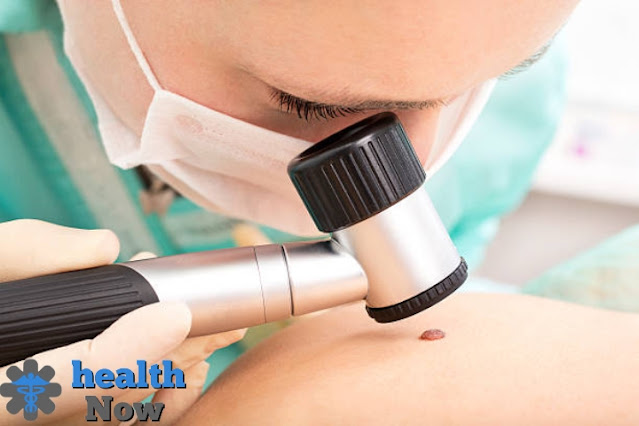
What is melanoma?
melanoma cancer is one of the most common types of skin cancer. It is also the most common form of cancer in men and the second most common in women.
It is often found in areas of the skin that are exposed to the sun, such as the skin on the head, neck, arms, and legs.
It can also occur in areas of the skin that are not exposed to the sun, such as the skin on the back, shoulders, and legs.
Melanoma is a type of skin cancer that originates in pigment-producing cells called melanocytes.
Melanoma is the most serious type of skin cancer and the most common primary cancer in males and females combined.
Melanoma is also the second most common type of cancer in men and the third most common primary cancer in women, according to the American Cancer Society.
Although melanoma is the most dangerous type of skin cancer, it is also the least common.
Symptoms of melanoma.
Melanomas from anywhere in the body. It usually forms in areas exposed to the sun, such as the back, legs, arms, and face.
Melanomas may form in areas that don't get many suns, such as the soles of the feet, the palms of the hands, and the layers around the nails. These melanomas are more common in people with darker skin.
The principal signs and side effects of melanoma are for the most part:
A change in an existing mole.
- The development of new pigmented or unusually shaped growths on the skin.
- Melanoma does not always begin as a mole. It may also occur in otherwise normal skin.
Natural moles.
Normal moles are usually uniform in color — such as tan, brown or black — with a distinct border separating the mole from the surrounding skin.
They are usually oval or round and are usually less than a quarter-inch (about 6 mm) in diameter - the size of a pencil eraser.
Most moles begin to appear in childhood and new moles may form until about age 40.
Upon reaching puberty, most people have between 10 and 40 moles. Moles may change shape over time, and some may disappear with age.
Abnormal moles indicate the presence of melanoma.
To help you identify features of abnormal moles that may indicate the presence of melanomas or other skin cancers, remember the letters ABCDE:
- A for the asymmetric shape. Look for irregularly shaped moles, such as halves, that are very different in shape from each other.
- B for irregular edges. Look for moles with irregular edges, notched or rounded edges — which are characteristic of melanomas.
- C for color changes. Look for growths that have too many colors or an uneven distribution of colors.
- D for width. Search for new development in a mole that is bigger than a fourth of an inch (around 6 mm).
- E for advancement. Search for changes over the long run, for example, moles that fill in size or change in variety or shape. Moles may likewise foster new signs and side effects, like late tingling or dying.
Cancerous (malignant) moles can vary greatly in appearance. Some may show all of the changes listed above, while some may have only one or two abnormal features.
Invisible melanomas.
Melanomas may also appear on areas of the body that get little or no sun, such as the space between the toes and on the palm, the soles of the feet, the scalp, or the genitals.
They are sometimes referred to as discreet melanomas because they occur in places most people wouldn't think to check.
When melanoma occurs in people with darker skin, it often occurs in an area that is not visible.
Non-visible melanomas include:
- Melanoma under a nail. Petechial melanoma is a rare form of melanoma that occurs under the fingernail or toenail.
- It may also be found on the palms of the hands or the soles of the feet. It is more common in people of Asian descent, black people, and others with darker skin tones.
- Melanoma in the mouth, digestive tract, urinary tract, or vagina. Mucosal melanoma occurs in the mucous membrane that lines the nose, mouth, esophagus, anus, urinary system, and vagina.
- Mucinous melanomas are particularly difficult to detect because they are easily confused with more common conditions.
- Melanoma in the eye. Melanoma of the eye, also called ocular melanoma, usually occurs in the uvea — the layer below the white of the eye (sclera). Melanoma in the eye can cause vision changes and may be diagnosed during an eye exam.
Causes of melanoma.
A patient develops melanoma when the melanin-producing cells (melanocytes) that give the skin its color malfunction.
Skin cells foster in a precise and controlled way under ordinary circumstances. New, sound cells push old cells toward the outer layer of your skin, where they at last pass on and tumble off.
However, when the DNA of certain cells is harmed, new cells might start to outgrow control and at last structure a mass o malignant cells.
It is as yet not satisfactory what causes DNA harm in skin cells and how this prompts melanoma. Melanoma might be brought about by a mix of ecological and hereditary variables.
However, doctors believe that exposure to ultraviolet radiation from the sun and tanning beds and lamps is the main cause of melanoma.
UV rays do not cause all melanomas, especially those that affect areas of your body that are not exposed to sunlight.
This indicates that other factors may contribute to your risk of developing melanoma.
risk factors.
- light skin; Having less pigment (melanin) in your skin means less UV protection. If you have blonde or red hair, have light eyes, freckles, and sunburn easily, then you are more likely to develop melanoma than people with darker skin. However, melanoma can also affect people with darker skin, including those with dark skin and Hispanic skin.
- History of sunburn. Having one or more sunburns with blistering can increase your risk of developing melanoma (melanoma).
- Excessive exposure to ultraviolet (UV) rays. Exposure to ultraviolet rays from the sun, lamps, and tanning beds can increase the chances of developing skin cancers such as melanoma.
- Living near the equator or in hot regions. People living near the equator in places where the sun's rays directly hit are exposed to greater amounts of ultraviolet rays than those living north or south of it. If you also live at an altitude, you will be exposed to more UV rays.
- Having a lot of moles or unusual moles. Having more than 50 normal moles on your body indicates an increased risk of melanoma (melanoma). Also, having an unusual type of mole increases your risk of developing melanoma (melanoma). Medically known as dysplastic birthmarks, moles tend to grow larger than normal, have irregular edges and have a mix of colors.
- Family history of melanoma (melanoma). If a close family member — such as a parent, son, or brother — has had melanoma (melanoma), you're more likely to develop it as well.
- Weakened immune system. People with compromised immune systems have an increased risk of melanoma and skin cancers. Your immune system may be damaged if you take medication to suppress your immune system, such as after organ transplants, or if you have a disease that weakens your immune system, such as acquired immunodeficiency syndrome.
protection.
You can reduce your risk of developing melanoma and other types of skin cancer by:
- Keep away from openness to the sun during the early afternoon time frame. For some individuals in North America, the sun's beams are most grounded between 10 a.m. furthermore, 4 p.m. So plan open air exercises at different times, even in winter or when the sky is cloudy.
- You absorb UV rays all year round, and clouds provide little protection from harmful rays. Avoiding the sun's rays when it's most intense can help you avoid sunburns and suntans that cause skin damage and increase the risk of skin cancer. Cumulatively, over time, sun exposure may also cause skin cancer.
- Use sunscreen all year round. Use a broad-spectrum sunscreen with an SPF of at least 30, even on cloudy days. Apply generous sunscreen and apply every two hours — or more often when swimming or sweating.
- Wear protective clothing. Cover your skin with dark, tightly woven clothing to cover your arms and legs, and wear a wide-brimmed hat; This provides more protection than a baseball cap or a face cap.
- Some companies also sell protective clothing. A dermatologist can recommend an appropriate brand. Don't forget to wear sunglasses. Look for ones that block both types of UV rays — UVA and UVB rays.
- Avoid burgundy lamps and beds. UV rays are emitted by russet lamps and harvesting beds and can increase your risk of skin cancer.
- Be aware of your skin characteristics so you can notice changes. Check your skin from time to time for new skin growths or any changes in existing moles, freckles, bumps, and birthmarks. Use mirrors to check your face, neck, ears, and scalp.
- Examine your chest, torso, and top and bottom of your arms and hands. Examine both the front and back of your legs and feet, including the soles of your feet and the spaces between your toes. Also, examine the genital area and between the butts.
Melanoma Diagnosis.
- Physical examination. Your doctor will ask you questions about your health history and examine your skin for signs that may indicate melanoma.
- Take an example of tissue for assessment (biopsy).To decide if a thought skin injury is a melanoma, your PCP might suggest a skin biopsy for testing. The example is shipped off to the research center for assessment.
- The type of biopsy your doctor recommends depends on your particular situation. Most often, doctors recommend removing the entire growing portion when possible. One common technique, stinger biopsy, is performed using a circular blade that is pressed into the skin around a suspicious lump.
- Another technique. called an excisional biopsy, which uses a scalpel to cut out the entire tumor mass and a small portion of healthy tissue around it.
Determining the extent of melanoma.
- Decide the thickness. The thickness of the still is up in the air via cautiously looking at the melanoma under a magnifying lens and estimating it with an extraordinary instrument. The thickness of the melanoma assists specialists in deciding on a treatment plan. As a general rule, the thicker the growth, the more serious the illness.
- More slender melanomas may just expect a medical procedure to eliminate the malignant growth and some typical tissue around it. On the off chance that the melanoma is thicker, your primary care physician might prescribe extra tests to check whether the disease has spread, before settling on treatment choices.
- Check whether the melanoma has spread to the lymph hubs. On the off chance that there's a gamble of disease spreading to your lymph hubs, your primary care physician might suggest a method known as a sentinel hub biopsy.
- During a sentinel node biopsy, a dye is injected into the area where the melanoma was removed. The dye flows to nearby lymph nodes. The first lymph nodes that absorb the dye are removed and tested for cancer cells.
- If these first lymph nodes (sentinel lymph nodes) are free of cancer, there's a good chance the melanoma hasn't spread beyond the area where it was first discovered.
- Look for signs of cancer outside the skin. For people with more advanced melanomas, doctors may recommend imaging tests to look for signs of cancer spreading to other areas of the body.
- Imaging tests may include x-rays, computed tomography, and positron emission tomography (PET) scans. These imaging tests are generally not recommended for smaller melanomas with a low risk of spreading beyond the skin.
Other factors may determine the risk of cancer spreading (metastasis), including whether or not the skin over the area has formed an open sore (ulceration) and the number of dividing cancer cells (mitosis) that are found when viewed under a microscope.
The stage of melanoma is determined using the Roman numerals 0 (zero) to IV. In stage 0 and stage I, the melanoma is small and the cure rate is very successful.
Yet, the higher the number, the lower the possibilities of full recuperation. In stage IV, the disease has spread externally the skin to different organs, like the lungs or liver.
Treatment.
The best treatment for melanoma depends on the size and stage of cancer, as well as your overall health and personal preferences.
Small melanoma treatment.
Treatment of melanoma in its early stages usually requires surgery to remove it. Very thin melanoma can be completely removed during a biopsy and does not require further treatment.
In other cases, however, the surgeon removes the er with a surrounding portion of healthy skin and a layer of subcutaneous tissue. This may be the only treatment needed for people with early-stage melanoma.
Treatment of subcutaneous melanoma cases.
- Surgery to remove affected lymph nodes. Assuming the melanoma has spread to the lymph hubs, the specialist might eliminate the impacted hubs. Extra medicines may likewise be suggested previously or after a medical procedure.
- Immunotherapy. Immunotherapy is a medication therapy that assists your resistant framework with battling disease. Malignant growth may not be gone after by your body's invulnerable framework, which is answerable for battling illnesses.
This is because disease cells produce proteins that they stow away from cells of the safe framework. Immunotherapy works by disrupting that interaction.
Immunotherapy is often recommended after surgery to remove melanoma that has spread to lymph nodes or other places in the body. When surgery can't completely remove the melanoma, immunotherapies may be injected directly into the melanoma.
- Targeted therapy. Targeted drug therapies focus on specific weaknesses found within cancer cells. Targeted drug therapies by targeting these vulnerabilities can cause cancer cells to die. Cells from melanoma can be tested to determine whether targeted therapy is likely to be effective in fighting cancer.
In the case of melanoma, targeted therapy may be recommended if cancer has spread to your lymph nodes or other areas of your body.
- Radiation therapy. This therapy utilizes high-energy radiates, like X-beams and protons, to kill disease cells. Radiation treatment might be coordinated to the lymph hubs on the off chance that the melanoma has spread. Radiation treatment may likewise be utilized to treat melanomas that can't be eliminated carefully.
Radiation therapy can help relieve symptoms if the melanoma has spread to other areas of the body.
- Chemotherapy. Chemotherapy uses drugs to kill cancer cells. Chemotherapy may be given intravenously, as a pill, or both ways to reach all parts of your body.
It may also be given through a vein in an arm or leg in a procedure called isolated perfusion of a limb.
In this procedure, blood in the arm or leg is not allowed to reach other areas of the body for a short time so that the chemotherapy drugs travel directly to the area around the melanoma without affecting other parts of your body.


