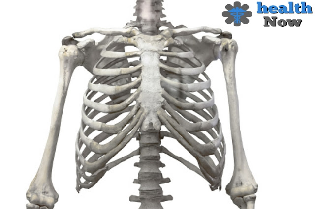
Rib cage.
The rib cage is part of the body's skeleton. The rib cage forms the human chest and protects important organs of life, namely the heart and lungs.
It consists of 12 pairs of ribs with a set of rib cartilage as well as a sternum, which also consists of several parts to be remembered later, and the ribs twist back to connect to the spine using pectoral vertebrae 1-12, which provides stability.
The ribs can be divided into groups. The first group is the real ribs and includes ribs Nos. 1 to 7, after which the ribs come from 8 to 12 and are called false ribs. In the last group of ribs, the last couple has a special name: floating ribs.
rib cage site.
The rib cage is the bone frame of the pectoral wall. The components of the rib cage are linked with each other firmly from the back to the vertebrae of the spine. From there, the ribs begin to twist and head forward, enclosing the pectoral cavity. The pectoral cavity is divided into three sections:
- The medium chamber containing the main cardiovascular
- And two side stones of the lungs.
The rib cage also connects to the neck from the top and supports the upper limbs, and from the bottom separates the chest from the abdomen by the respiratory diaphragm muscle.
Rib cage parts.
Now, after forming a general picture of the rib cage, its shape, and its potential, it is necessary to look in more detail at its parts in detail, every part also consists of other parts that are more important to identify more precisely.
As follows: 0 seconds of 0 seconds.
Shear bone.
It is the center of the rib cage and its fixation from the front, a long bone consisting of three parts, the first part is known as the manubrium shear grip which is the upper and wider part of the shear bone, and the upper edge of the sheer grip can be easily and felt.
It is the shallow, U-shaped bone at the front base of the neck and between the collarbone, known as the overstepping bone, and the first two ribs are linked to the shear fist, after which the second part, the central and extended part of the cloth, is called the shear body, where it is attached to the fist by the poem angle.
It's called because it's a little curved and not flat, and at this angle, it binds the 2 ribs, the first and highest that can be felt, because the first two ribs disappear behind the collarbone, and the shear angle is a distinctive feature of the 3-7 rib calculation attached to the shear body.
Finally, the lower end comes called the sword excess or monastic cantilever xiphoid process, a small part that is cartilage at the beginning of life, and in middle age, it becomes gradually solid.
ribs.
The rib cage contains 12 pairs of ribs. Each rib is a flat and curved bone that contributes to the construction of the rib cage wall. The ribs extend backward to meet and relate to the chest spine vertebrae from the first paragraph to paragraph 12. Depending on the numbering of the spine's vertebrae, the ribs carry the same numbering from 1 to 12.
From the front, each rib is linked to the sternum through its rib cartilage, as well as to cartilage several parts. The rear end of the rib is called the rib head. Besides the head, there is a narrow rib neck. On the back surface of the rib, there is a small bump called the rib canine, where it meets and accidentally separates from the paragraph bearing.
Aligned with the angle of the rib, which is the point with the greatest degree of curvature, the angles of the ribs line the middle of the shoulder. The remainder of the rib, which is the axis or extended part, is the body of the rib. There is a shallow corridor or channel along the lower edge of each rib for the passage of nerves and blood vessels.
The ribs are divided into three types, the first being the real ribs, the first seven ribs, and their rib cartilage is directly associated with the sternum, after which the false ribs come from the 8 to the 12 and their cartilage is not directly related to the sternum, where the 8-10 rib cartilage is directly related to the rib that precedes them.
In other words, rib cartilage 10 is associated with rib cartilage 9 and so on, and ribs 11 and 12 have another name as well: floating ribs, two short ribs that are not related to the cut, but their cartilage ends in the side muscles of the abdominal wall.
Rib cartilage.
It is the cartilage that binds the ribs to the sternum and helps them to expand and move forward. It is the presence of this cartilage that gives flexibility to the chest walls that is necessary for its expansion during the breathing process. Since the number of ribs is 12, the cartilage is also 12 cartilage.
Each has two cartilage in the form of frontiers and limbs, with seven pairs associated with the sternum, three pairs associated with the rib before it, and the two ear ribs ending in the abdominal wall are hot and have two sharp cartilage.
The cartilage has a convex front surface, the rear is concave, the upper boundaries are convex and the bottom is concave, and despite that flexibility, with age and reaching 65
Age becomes prone to sclerosis and fossilization.
Spaces between ribs.
After identifying the shape of the rib cage and how to arrange ribs and cartilage, it becomes clear that this structure results in spaces or spaces between them called the spaces between ribs, and there are 11 spaces between ribs.
These spaces separate each rib, its cartilage, and the other rib, moving the rib cage during breathing smoother and more flexible. These spaces also bear names. These spaces are called according to the cartilage that forms the upper limit.
For example, the area between ribs 4 and 5 bears the number 4, and these areas contain muscles and membranes, 11 nerves between ribs, and two sets of blood vessels with the same rib area number.
joints.
A general picture of the body of the rib cage is formed with its parts and components. Until the picture is complete, we should talk about the last parts of the rib cage, which are the joints. The rib cage includes many joints, as follows:
- The joints that separate the vertebrae of the spine.
- The joints separate the sternum bone from the collarbone.
- The joints between the sternum and the rib cartilage 1-7.
- The joint between the bone of the shear grip and the sheer body.
- The joint between the shear body and the sword excess.
- The joints between the ribs and the rib cartilage.
- The joints between the ribs and the corresponding paragraphs.
- The joints separate the rib cartilage.
Functions of the rib cage.
Because the rib cage is part of the skeleton and depending on its location and composition, it protects all organs and internal ingredients in the chest area, most importantly the lungs and heart, and can do so.
The rib cage should have flexibility in movement and protection, and this flexibility is already present thanks to the small joints that link the ribs to the spine but it does not mean freedom of movement but is limited in proportion to the functions required.
The functions of the rib cage are not limited to protecting against any external force, as the upper limbs of the body attached to it provide support and stability, and it is the center of the attachment and stabilization of many muscles responsible for controlling the movement and direction of the upper limbs and coordinating them with the torso.
The most important function of the rib cage is its important role in the breathing process. The rib cage is semi-rigid and capable of expanding, due to several muscles, the most important of which is the diaphragm separating the chest from the abdomen, as well as the fibrous ligaments and tendons between it and the chest vertebrae in the spine.
When the rib cage expands, the air pressure within the lungs decreases compared to the outside air, pushing the air to move and enter the lungs to rebalance the pressure, which is the inhalation process.
The process of exhaling is reversed when muscles and ligaments, including the diaphragm, relax, and the composition of the last five ribs gives the lower part of the rib cage and diaphragm freedom of movement and breadth.
Diseases of the rib cage.
my asylum comes out or the rib cage pops up.
One of the diseases of the rib cage is the impulse of the pectoral bone and its prominence outside its natural position in the chest, also called the chest of the pigeon, and infected with this disease among 1,500 children, it is the second most abnormal condition affecting children.
Males are four times more likely to be infected than females. This condition usually starts from childhood and during the development and development of cartilage that binds the ribs. Instead of taking their natural shape, they appear outside, accompanied by a range of symptoms, including:
- Accelerated heartbeat.
- Chest pains.
- Shortness of breath, especially when doing exercises.
- Feeling tired.
- Recurrence of respiratory infections.
- Having a crisis or asthma.
- Pain in the area of abnormal growth of cartilage.
There are two types of the disease, the first type is called tis the chicken's chest, which highlights the median and lower part of the chest bone and is the most prevalent.
The second type, the pigeon's chest, highlights the upper part of the bone, forming a Z-like, and the real cause of the condition is unknown but results from the abnormal evolution of cartilage.
It has the function of pressing the affected area to adjust it, and this treatment benefits in pre-puberty, and then the doctor resorts to surgery.
broken ribs of the rib cage.
When looking for the most health problems or diseases affecting the rib cage, rib fractures are in the first position, cracking or breaking rib bones. The main reason for this is usually a strong blow to the chest as a result of falls, or car accidents, for example.
A rib fracture is often only a fracture, and although it causes pain, it remains unrisky, such as in cases of fracture and separation of broken parts, because this condition can result in significant damage to major blood vessels and important organs such as the lungs due to the sharp limbs of the broken bone, and in most cases, the ribs are self-healed within a month or two.
But it is necessary to control pain until healing, the symptoms of fractures are pain that increases when taking a deep breath, pressing the place of injury, or trying to bend and twist the body.
Many factors increase the chance of such fractures. Osteoporosis and osteoporosis make bone fractures easier. Practitioners of certain types of sports such as footballers and hockey players are often exposed to falls or chest blows, and a wound or cancer injury weakens the structure of the bones.
Therefore, the necessary precaution and prevention methods must be taken as athletes' clothing for proper protectors, relieving the triggers of slipping and falling inside the house such as fixing carpets, cleaning any spots that cause slipping and using a rubber carpet during the shower, and most importantly, ensuring that the bones are strengthened by taking the calcium and vitamin D necessary for bone strength.
Anterior cartilage inflammation.
Like any other part of the body, the rib cartilage in the rib cage may be exposed to inflammation. This type of inflammation usually affects the cartilage that connects the upper ribs to the sternum.
Inflammation is accompanied by chest pain ranging from mild to sometimes very severe. In mild cases, pain may be limited to pressure on the inflammation area.
Severe cases of pain are severe and unlikely, to affect a person's life. Pain can extend to all ribs. Inflammation occurs as a result of some types of joint infections, tumors in the area of the rib cartilage, some respiratory diseases, and viruses such as tuberculosis and syphilis.
After diagnosis, many therapeutic procedures are followed. Most cases of anterior cartilage are treated with non-steroidal anti-inflammatories such as ibuprofen and naproxen. Some tricyclic antidepressants can be prescribed as well as some types of steroids. Lifestyle should be modified in the lying and resting person, using hot or cold compresses.
Tetz syndrome.
A rare disease that affects the musculoskeletal system, very painful but not serious, occurs when the cartilage surrounding the upper rib bone is swollen, and the second or third rib is usually the most infected, and scientists have not been able to find the cause of this syndrome, but they are likely to suffer mild blows to the chest wall.
The condition can also affect patients who have had a lot of respiratory infections, and those under 40 years of age are more likely to develop Titz syndrome. The most characteristic of this condition is swelling as well as chest pain, where pain appears and then suddenly disappears.
Or the pain may graduate and appear and then disappear over years, and the pain may disappear, but the swelling persists, the intensity and intensity of the pain varies, it may be mild or resemble stabbing with a knife, and some may think it is heart attack pain, as it may extend to the neck of the shoulders and arms.
Some movements can aggravate pain such as sneezing and coughing, breathing deep, laughing, and tying the seatbelt when riding, and their treatment is simple in the use of non-steroidal antiinflammants and can also use steroids.
Padgett's disease.
This disease occurs as a result of a local bone formation disorder, which begins with excessive osteoporosis and then is followed by a large activity in the formation and formation of the bone, resulting in this disturbance in bone cell activity creating an irregular mosaic bone that makes it weaker, larger.
Less pressure, more vascular, and easier to break as well. Most sufferers may not show any symptoms. However, symptoms are osteoporosis, neurological problems, excessive warmth, and secondary osteoporosis as well as osteoporosis, which affects different parts, but the axial skeleton is the most prone.
You can stay without a rib cage.
To answer this question, it is necessary to mention a medical condition dealt with by doctors, a condition of a 76-year-old man.
He lived a decade of life without his sternum, suffering from diseases in the coronary arteries and vascular system, undergoing coronary artery conversion, and the process was complicated by inflammations that led to sternites, necessitating the partial removal of the sternum and ribs.
As a result, the front chest skeleton has become incomplete, and parts of the heart and lungs have been exposed and unprotected for more than 10 years.


